Presbyterian Heart & Vascular Care Providers
Our team of highly skilled doctors and clinicians offers a full range of heart-related services from diagnosis and treatment to monitoring.
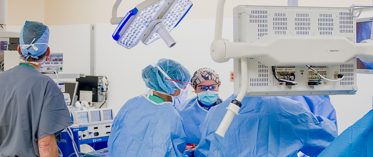
We have a highly skilled team who can provide a wide range of services from diagnosis to treatment.
Congenital heart disease occurs while a fetus is developing. It's seen in 1% of babies born in the U.S. and is the most common birth defect.
The fetal heart is completely formed by eight weeks. After that, a fetal and perinatal echocardiogram can use sound waves to create pictures of an unborn baby's heart and detect any heart defects.
A specially trained ultrasound technician typically performs the test, and a pediatric cardiologist (heart doctor) interprets the images.
Heart defects range from mild to very severe. Some require surgery for newborns and some get better on their own.
Your obstetrician or midwife will talk with you about fetal or perinatal echocardiography if they think one is needed.
Some women are more at risk of having a baby with a heart defect. Things that make it more risky include:
Fetal and perinatal echocardiography can show heart disease or heart defects.
A congenital heart defect is a structural problem of the heart that develops during pregnancy. About 1/100 babies are born with a heart defect. Most people with these defects are treated when they are newborns.
It is always important to have as much information as possible when you get a fetal echocardiogram. You'll want to know why your obstetrician or perinatologist wanted you to have one. If the reason is that you have a heart defect, bring your medical records.
An ultrasound technician will do the echocardiogram and a pediatric cardiologist (children's heart doctor) will review the pictures.
There are two ways to do this test:
You will have the test in a dark room while lying down. It is like a regular ultrasound. You will feel some pressure from the wand, but no pain.
It can take 30 minutes to two hours to get the pictures needed to see all the parts of the heart.
Presbyterian’s Pediatric and Congenital Cardiology team has many different options to help you manage your or your child’s heart condition. The team performs various diagnostic tests and procedures to help form an accurate diagnosis and create individualized treatment plans. Depending on the type of heart condition your child has and its underlying cause, the team can recommend a wide variety of treatment options. Our pediatric cardiologists, pediatric interventional cardiologists, and pediatric cardiovascular surgeons work closely together for cases in which cardiac repair or surgery is the best treatment option.

Our team of highly skilled doctors and clinicians offers a full range of heart-related services from diagnosis and treatment to monitoring.